2.1. VLPFC (area 45a)
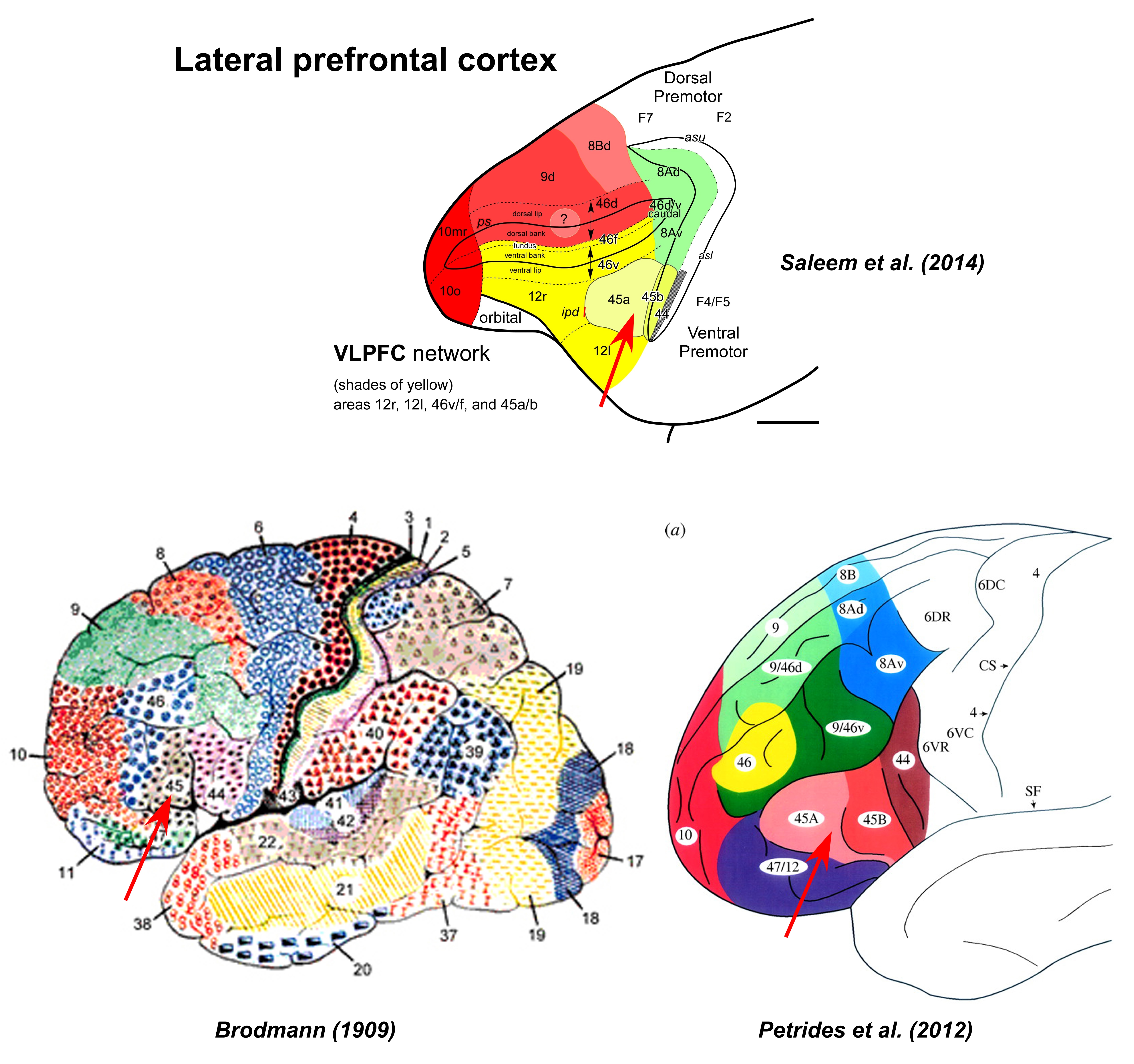
VLPFC (area 45a) Area 45 is found in the caudal aspect of the convexity, back to the inferior limb of the arcuate sulcus ( Walker, 1940 ). Based on the comparative cytoarchitectonic analysis in both human and nonhuman primates, Petrides and Pandya (1999, 2002) have subdivided this area into 45a and 45b, on the convexity rostral to the arcuate sulcus and in the rostroventral bank of the sulcus, respectively (see also Gerbella et al., 2010, for the architectonic analysis of these areas ).
2.1.1. Images
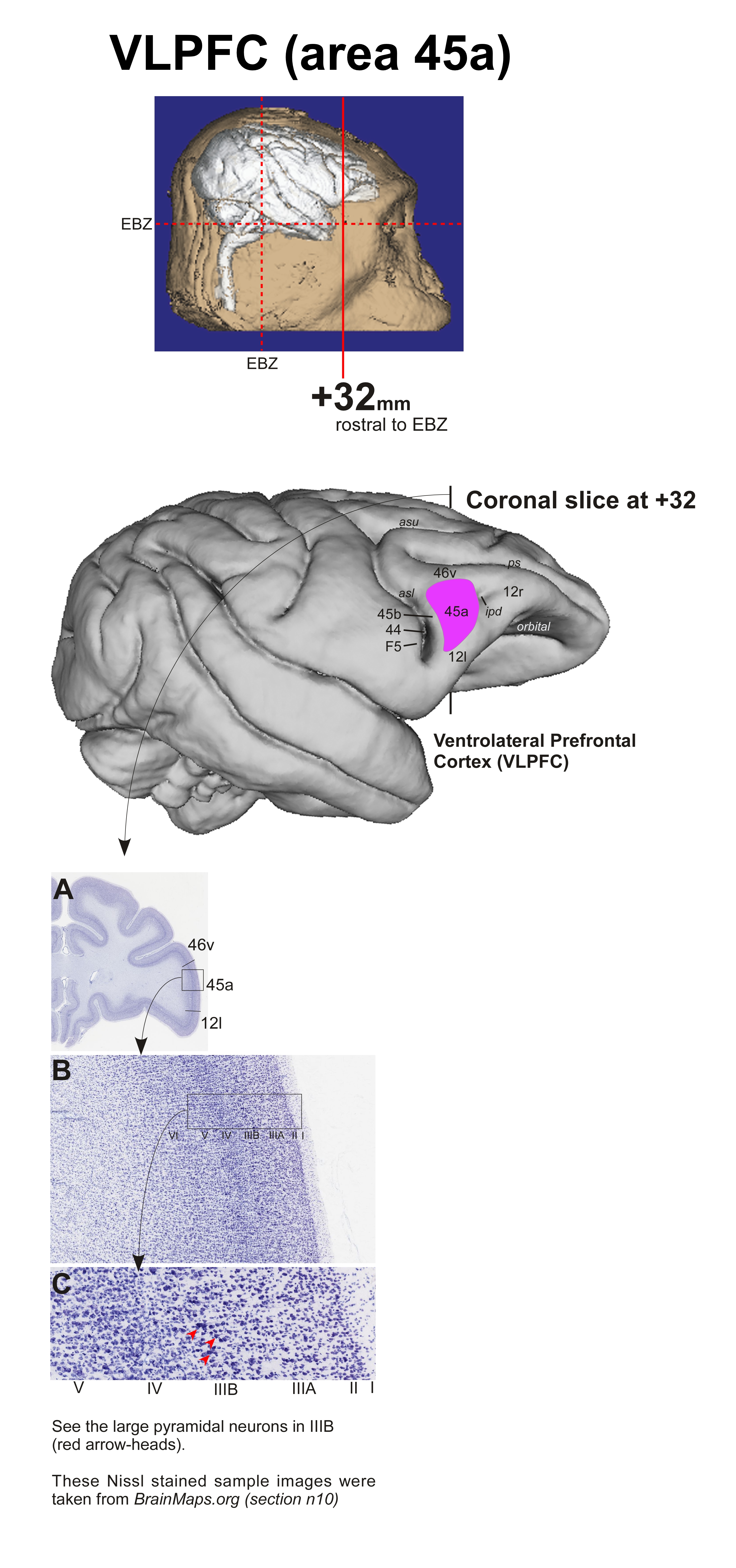
area 45a histology sections courtesy of brainmaps.org
2.1.2. Architectonic delineation
Area 45a is characterized by the presence of large but less prominent pyramidal neurons in deeper part of layer III (IIIB; see red arrow-heads in Fig. C on the left); poorly defined layer II; well developed layer IV; and densely populated small and medium sized pyramidal neurons in layer V (See also Walker, 1940; Petrides and Pandya, 1999, 2002; Gerbella et al., 2010; Petrides et al., 2012 ).
2.1.3. Connections
Find more info on Area 45a connections at the following link
| Type | Connections |
|---|---|
| Intrinsic | Area 45a is connected with all the subdivisions of the LPFC (VLPFC, DPFC and CLPFC) networks. |
| Extrinsic | The major connections are with auditory related areas in the supratemporal plane (STP), superior temporal gyrus (core, belt, parabelt, and STGr), and the dorsal bank of the superior temporal sulcus (STSd; Petrides and Pandya 2002; Saleem et al., 2008, 2014; Gerbella et al., 2010 ). |
| Subcortical |
45a also receives feedback projections from magnocellular, parvicelluar, and intermediate subdivisions of the basal nucleus (Bmc, Bpc, Bi; Gerbella et al., 2013 ). |
2.1.4. Functions
- Vocal and facial communication between monkeys.
- Recordings that seem to correspond to 45a have found convergent of visual and auditory stimuli in individual neurons; the auditory responses were particularly to species-specific calls (Sugihara et al., 2006; Romanski, 2012 ).
- Visual responses to face stimuli have also been found ( Wilson et al., 1993; O’Scalaidhe et al., 1997; Tsao et al., 2008 ).
2.1.5. Homologue area in human
Area 45a (also 45b and 44) may be a precursor for the language related areas found in the caudal ventrolateral prefrontal cortex in humans ( Kelly et al., 2010; Petrides et al., 2012, 2014 ).
area 45a macaque human comparison
2.1.6. References
Contini M, Baccarini M, Borra E, Gerbella M, Rozzi S, Luppino G. 2010. Thalamic projections to the macaque caudal ventrolateral prefrontal areas 45A and 45B. Eur J Neurosci 32:1337-1353, 2010. PubLink
Ferry AT, Ongur D, An X, Price JL. 2000. Prefrontal cortical projections to the striatum in macaque monkeys: evidence for an organization related to prefrontal networks. J Comp Neurol 425:447-470. PubLink
Gerbella M, Baccarini M, Borra E, Rozzi S, Luppino G. 2013. Amygdalar connections of the macaque areas 45A and 45B. Brain Struct Funct. Mar 26. PubLink
Gerbella M, Belmalih A, Borra E, Rozzi S, Luppino G. 2010. Cortical connections of the macaque caudal ventrolateral prefrontal areas 45A and 45B. Cereb Cortex 20:141-168. PubLink
Kelly C, Uddin LQ, Shehzad Z, Margulies DS, Castellanos FX, Milham MP, Petrides M. 2010. Broca’s region: linking human brain functional connectivity data and non-human primate tracing anatomy studies. Eur J Neurosci 32:383-398. PubLink
O’Scalaidhe SP, Wilson FA, Goldman-Rakic PS. 1997. Areal segregation of face-processing neurons in prefrontal cortex. Science 278:1135-1138. Petrides M. 2014. Neuroanatomy of language regions of the human brain. Academic Press, San Diego. PubLink
Petrides M, Pandya DN. 1999. Dorsolateral prefrontal cortex: comparative cytoarchitectonic analysis in the human and the macaque brain and corticocortical connection patterns. Eur J Neurosci 11:1011-1036. PubLink
Petrides M, Pandya DN. 2002. Comparative cytoarchitectonic analysis of the human and the macaque ventrolateral prefrontal cortex and corticocortical connection patterns in the monkey. Eur J Neurosci 16:291-310. PubLink
Petrides M, Tomaiuolo F, Yeterian EH, Pandya DN. 2012. The prefrontal cortex: comparative architectonic organization in the human and the macaque monkey brains. Cortex 48:46-57. PubLink
Romanski LM. 2012. Integration of faces and vocalizations in ventral prefrontal cortex: implications for the evolution of audiovisual speech. Proc Natl Acad Sci U S A 109:10717-10724. PubLink
Saleem KS, Kondo H, Price JL. 2008. Complementary circuits connecting the orbital and medial prefrontal networks with the temporal, insular, and opercular cortex in the macaque monkey. J Comp Neurol 506:659-693. PubLink
Saleem KS, Miller B, Price JL. 2013. Subdivisions and connectional networks of the lateral prefrontal cortex in the macaque monkey. J Comp Neurol Nov 9 [Epub ahead of print]. PubLink
Sugihara T, Diltz MD, Averbeck BB, Romanski LM. 2006. Integration of auditory and visual communication information in the primate ventrolateral prefrontal cortex. J Neurosci 26:11138-11147. PubLink
Tsao DY, Schweers N, Moeller S, Freiwald WA. 2008. Patches of face selective cortex in the macaque frontal lobe. Nat Neurosci 11:877-879. PubLink
Walker AE. 1940. A cytoarchitectural study of the prefrontal area of the macaque monkey. J. Comp Neurol 73:59-86. PubLink
Wilson FA, Scalaidhe SP, Goldman-Rakic PS. 1993. Dissociation of object and spatial processing domains in primate prefrontal cortex. Science 260:1955-1958. PubLink