Hi!
I am running skullstripping on a large (>2000) T1w images from different sites and hence different image parameters. I started using the FSL bet function since we plan on using the FSL VMB function package later on and I found that for some datasets there is always a quite large chunk of tissue left form the area that is between the brain and spinal cord (left of the CSF that is left of the Pons).
I changed to AFNI's 3dSkullstrip and I have the same problem. I played around with some of the tips in the 3dSkullstrip function description but those are mostly for when GM is missing.
Usually this is no problem for fMRI analysis since the T1 is warped and then masked but I want to use these T1w images in native space for some Deep learning applications.
Do you have any tips on how to improve the skullstrip for these datasets?
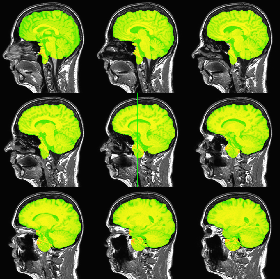
See attached image. Thanks in advance!


